
Hacettepe University, which brings together academic knowledge and research, is increasing the use of domestically developed technology in the healthcare sector for the benefit of patients. The domestically developed ‘digital twin’ system, created within our university’s Neurosurgery Education, Research and Innovation Centre (HÜNERİM), enables the planning of high-risk and complex surgeries in a virtual environment. The system enables the three-dimensional modelling of the patient’s brain tissue, blood vessels, nerves and the affected area using radiological images.
The resulting three-dimensional model offers surgeons the opportunity to examine in detail the position of critical structures surrounding the surgical site, such as brain tissue, nerves and blood vessels. Using these models, the access route to be used during surgery can be determined, different surgical approaches can be assessed in advance, and the operation can be planned according to the patient’s own anatomy.
University and Technopark collaboration
Developed in collaboration with Hacettepe Technopark and named the ‘Personalised Surgical Anatomy Visualisation Platform (Persanavi)’, this technology also significantly reduces the time required to create 3D models. The modelling process, which used to take days, can now be completed in a matter of minutes with the support of artificial intelligence and computer technology.
It enables the training of better doctors
Prof. Dr Mustafa Berker, Head of the Department of Neurosurgery at Hacettepe University Faculty of Medicine, stated that the work carried out within the HÜNERİM programme contributes both to the development of patient-specific treatment methods and to neurosurgery training, and made the following statement:
“Through HÜNERİM, we are developing more advanced treatment methods using patient-specific treatment planning and innovative techniques. This not only ensures that patients receive advanced treatment but also contributes to the training of better doctors in the field of neurosurgery. Our primary aim is to provide the best possible education and, in this way, deliver the best possible healthcare.”
Prof. Dr Berker emphasised that the primary objective of the work is to provide patients with the best possible healthcare by enhancing the quality of education.
Three-dimensional models show the patient’s actual anatomy
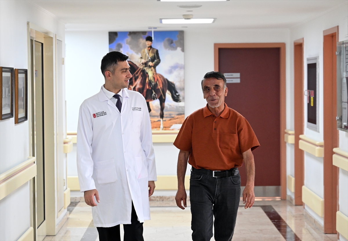
Associate Professor Dr Şahin Hanalioğlu, a lecturer in the Department of Neurosurgery at Hacettepe University Faculty of Medicine, noted that CT and MRI scans are among the fundamental tools for surgical planning, whilst pointing out that these methods provide two-dimensional cross-sectional images. Explaining that the anatomical structures in these images are transformed into realistic models using three-dimensional reconstruction and computer vision techniques, Hanalioğlu stated that artificial intelligence and computer technologies have significantly accelerated the process.
Pointing out that three-dimensional models present the patient’s actual anatomy in detail to the surgeon, Hanalioğlu said, “Three-dimensional models lay the patients’ actual anatomy out before our very eyes. In this respect, we are among the top 10 centres in the world and part of a team with this capability.”
The surgeon can assess different scenarios before the operation
Hanalioğlu, noting that the technology makes a significant contribution to surgical planning, particularly in complex cases, continued:
“Visualising the patient’s actual 3D anatomy in one’s mind is crucial. With this technology, the surgeon has the opportunity to directly observe the surgical scenario they will encounter during the operation. They can, in effect, carry out a digital ‘rehearsal’ of the operation they are due to perform the following day the day before. They can also make use of these 3D models during the operation.”
Stating that the technology can be used in the surgery of complex tumours and vascular diseases, Hanalioğlu noted that, thanks to this technology, the surgeon can determine the direction of approach, as well as the position and size of the bone window to be created in the skull, based on the patient’s anatomy. Hanalioğlu also emphasised that, thanks to this technology, the relationship between the tumour and the surrounding brain tissue, nerves and blood vessels that need to be protected can be assessed in detail prior to surgery.
Hanalioğlu reported that the technology has been used in over 200 patients to date, that pathologies have been correctly identified and modelled in almost all of them, and that successful surgical outcomes have been achieved.
The tumour, measuring approximately 5 centimetres, was removed via a small incision
According to information obtained from Hanalioğlu, in one of the cases where digital twin technology was used, the surgery to remove a tumour measuring approximately 5 centimetres—located at the base of the skull between the two eyes and the olfactory nerves—was planned using a three-dimensional model. Hanalioğlu stated that, despite the tumour’s size, the operation was carried out via a small incision made within the eyebrow. Explaining that the location of the bone cut and various surgical options were assessed on the digital twin prior to the operation, Hanalioğlu said that, following this preparation, the tumour could be completely removed through a very small opening. Hanalioğlu shared that the patient, who was discharged shortly after the operation, is continuing to lead a healthy life.
The approach to the vascular malformation in the brainstem was planned in advance
The domestically developed ‘digital twin’ technology, created by our university, was used to plan surgery for a 56-year-old patient with a progressively enlarging vascular malformation in the brainstem. Professor Dr Ahmet İlkay Işıkay, a specialist in Neurosurgery at our university, stated that three-dimensional modelling played a significant role in determining the approach to be taken to reach the vital vascular malformation in the brainstem.
Sharing that the patient, whose condition was deteriorating, could only walk with the support of two people prior to the operation, Işıkay noted that the patient was unable to open their eyelids, move their eyes or speak. Işıkay explained that the operation lasted approximately five hours and that the vascular malformation in the brainstem had been removed. He reported that the patient recovered rapidly following the operation and had largely regained his health; he can now walk unaided, open his eyelids and his speech has improved.
The risk of bleeding from the large tumour was also assessed prior to surgery
Prof. Dr Mehmet Erhan Türkoğlu, a specialist in Neurosurgery at Hacettepe University, also stated that they utilised three-dimensional modelling during the surgery to remove a large tumour located in the posterior fossa and originating from the meninges. In his remarks, Prof. Dr Türkoğlu said:
“Thanks to pre-operative planning, we were able to assess the extent of the tumour’s spread within the venous system and whether removing it from this area would cause significant bleeding. Consequently, we were able to obtain more detailed information about the patient and take certain precautions against potential complications. The operation lasted approximately 8 hours. We were able to observe virtually every detail we had planned pre-operatively during the operation itself. As a result of the operation, we were able to remove the entire tumour. We were also able to discharge our patient home in good health.”
“After the operation, I felt as though I’d been reborn”
Patients whose operations were planned using 3D modelling also shared their assessments of their recovery processes following treatment. Ece Prim, aged 58, who stated that her headaches, which had persisted for around 5–6 years, had gradually worsened over time and had recently been accompanied by vomiting, explained that a tumour measuring approximately 7 centimetres had been detected in her brain following her symptoms.
Pirim, who travelled to Ankara for treatment, said that 15 days had passed since her operation and that she had not taken any painkillers during that time. Describing her experience after the operation, Prim said: “After the operation, I felt as if I’d been reborn. I can do my own work, I can speak, I can move around. It’s been a gift of a new life for me.”
“I can smell again now”
Mehmet Çanayaz, a 52-year-old resident of the Doğubayazıt district of Ağrı, stated that he had been unable to smell for around 10 months, adding that he had experienced occasional unease and noticed changes in his behaviour. Çanayaz, who was found to have a tumour measuring approximately 5 centimetres in his brain following examinations, explained that he had sought treatment at Hacettepe University on the recommendation of a relative who had previously undergone surgery performed by Assoc. Prof. Dr Şahin Hanalioğlu.
Noting that approximately 13–14 days had passed since his operation and that he had been able to get back on his feet shortly afterwards, Çanayaz said, “I can smell again now. I feel very well. Tomorrow, God willing, I’m flying to Doğubayazıt. I’m going to be reunited with my children and grandchildren.”